
Small-angle X-ray scattering reveals characteristics of medieval human bones
October 4, 2025
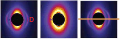
A new study, first-authored by graduate student Laura Gardner and published in Journal of the Royal Society Interface, demonstrates the capacity of small-angle X-ray scattering (SAXS) in the rapid and non-invasive assessment of collagen diagenesis in archeological bone. Second metacarpal bones of adult humans were obtained from a medieval cemetery in Bavaria, Germany, which was discovered during the construction of a new railway line. A Northwestern Feinberg School of Medicine researcher used a beamline of the Advanced Photon Source at Argonne National Laboratory to perform SAXS on the bones. The research team hypothesized that collagen diagenesis would be evident from the relatively low intensity of scattered light at a position in the SAXS patterns corresponding to the spacing of carbonated hydroxyapatite nanoplatelets within the bone -- spacing dictated by collagen fibrils. Conversely, they suspected that bones with well-preserved collagen would show high intensity peaks at the same position. The SAXS data was compared to the results of laboratory microcomputed tomography, microscopy, and Raman spectroscopy performed at DePaul University, the University of Hildesheim, and Northwestern DEEPS. In fact, the additional analyses validated the SAXS interpretations. The study illustrates how minerals like apatite are not only of geological significance, but are also important in archeological sleuthing.